What is Ultrasound ?
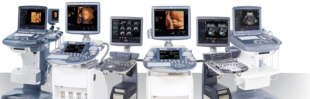
Ultrasound imaging uses sound waves to produce pictures of the inside of the body. It is used to help diagnose the causes of pain, swelling and infection in the body's internal organs and to examine a baby in pregnant women and the brain and hips in infants. It's also used to help guide biopsies, diagnose heart conditions, and assess damage after a heart attack. Ultrasound is safe, noninvasive, and does not use ionizing radiation.
This procedure requires little to no special preparation. Your doctor will instruct you on how to prepare, including whether you should refrain from eating or drinking beforehand. Leave jewelry at home and wear loose, comfortable clothing. You may be asked to wear a gown.
What is Color Doppler?
Color Doppler is a method of visually detecting motion or blood flow using a color map that is incorporated into a standard B-mode image. The principles of color Doppler are similar to those of pulsed-wave Doppler. However, a larger region can be interrogated, and detected blood flow is assigned a color, typically blue or red, depending on whether the flow is moving toward or away from the transducer. Frequency shifts are estimated at each point at which motion is detected within an interrogated region, thus yielding information on direction of motion and velocity. Shades of blue or red are used to reflect the relative velocities of the blood flow. All stationary objects are represented on a gray scale, as in B-mode imaging. The benefit of color Doppler is that information on the direction and relative velocity of blood flow can be obtained. Color Doppler is limited by its dependence on the relative angle of the transducer to the blood flow.